06/27/2019 11:00 AM EDT
The National Library of Medicine recently acquired the Patient/Problem Oriented Medical Record System Archives, a collection of materials related to the development of an early


 |
 |
 |
 |
 | |
Top Story3D printed technology streamlines common medical testScientists have taken a common, yet laborious lab test and redesigned it to be performed in small 3D printed pipette tips used to measure and transfer fluids in the laboratory. Read more... |

06/27/2019 | |
 | Footage of laboratories at the Advanced Technology Research Facility in Frederick, Maryland, part of the Frederick National Laboratory for Cancer Research. Included are: an LTQ Orbitap Velos mass spectrometer in use at the Laboratory of Proteomic and Analytical Technologies; a two-photon scanning microscope in use in the Optical Microscopy and Analysis Lab; a nuclear magnetic resonance spectrometer in use at the Laboratory of Proteomic and Analytical Technologies; a 1000- liter bioreactor in use at the Biopharmaceutical Development Program; a multi-touch table for a prototype of visual imaging collaboration environment at the Advanced Biomedical Computing Center; and a 200-kV transmission electron microscope in use at the Electron Microscopy Lab. |
06/27/2019 | |
 | Includes footage of mammography, digital mammography, Magnetic Resonance Imaging (MRI), palpation, sigmoidoscopy, digital rectal exam (DRE), chest X-ray, spiral CT, and pap test. |
06/27/2019 | |
 | Includes images of a man smoking, a man putting out a cigarette, a woman shopping for fruits and vegetables, and close-up images of fruits and vegetables. This video has no audio. |
06/27/2019 | |
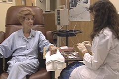 | Footage of chemotherapy and radiation. Shows chemotherapy bags, women in a chair receiving chemotherapy, a woman on a radiation table, and radiation equipment. This video has no audio. |
06/27/2019 | |
 | Includes lab footage, as well as footage of a patient getting a vaccine. This video has no audio. |
06/27/2019 | |
 | For the first time, scientists have directly observed events that lead to formation of a chromosome abnormality that is often found in cancer cells. The results of this study--from senior author Tom Mistelli, Ph.D., lead author Vassilis Roukos, Ph.D., and colleagues at the National Cancer Institute--appeared in the Aug 9, 2013, issue of the journal Science. |
06/27/2019 | |
 | On September 29-30, 2009, the U.S. National Cancer Institute, part of the National Institutes of Health, signed letters of intent to collaborate in cancer research efforts with the governments of Argentina, Brazil, Mexico and Uruguay. These bilateral partnerships aim to accelerate progress against cancer in Hispanic populations in the United States and Latin America and improve cancer research. |
06/27/2019 | |
 | Footage from the NCI Cancer Genomics Research Laboratory. Footage shows equipment used in the Genome-Wide Association Studies labs. This video has no audio. |
06/27/2019 | |
 | Footage from The Cancer Genome Atlas (TCGA), an NCI project co-sponsored by the National Human Genome Research Institute to map the cancer genome. Images include equipment, animation, and scientists. The beginning of this video has no audio. |
06/26/2019 | |
 | Anatomía de la columna vertebral. En el dibujo se observa una vista lateral de la columna vertebral, que incluye la región cervical (C1-C7), la región torácica (T1-T12), la región lumbar (L1-L5), la región sacra (S1-S5) y el cóccix (hueso coccígeo). También se muestran la médula espinal, las vertebras (huesos de la columna), el cono medular (parte final de la médula espinal), la cola de caballo (conjunto de nervios raquídeos que continúan más allá del cono medular) y los discos lumbares. Además, se muestra el clivus (hueso en la base del cráneo cerca de la médula espinal). |
06/26/2019 | |
 | Anatomía del aparato digestivo. En la imagen se observan la boca, las glándulas salivales, la faringe (garganta), el esófago, el estómago, el hígado, la vesícula biliar, el páncreas, el intestino delgado, el intestino grueso, el recto y el ano. |
06/26/2019 | |
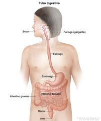 | Anatomía del tubo digestivo. En la imagen se observan la boca, la faringe (garganta), el esófago, el estómago, el intestino delgado, el intestino grueso, el recto y el ano. |
06/26/2019 | |
 | Glands and organs of the endocrine system; drawing shows the hypothalamus, pituitary gland, pineal gland, thyroid gland, thymus, adrenal gland, pancreas, ovaries (female), and testes (male). An inset shows the back view of the thyroid gland with the four pea-sized parathyroid glands on it. |
06/26/2019 | |
 | Two-panel drawing of stage IB lung cancer; the panel on the left shows a tumor (larger than 3 cm but not larger than 4 cm) in the right lung. Also shown are the pleura and diaphragm. The panel on the right shows a primary tumor (4 cm or smaller) in the left lung and cancer in (a) the left main bronchus and (b) the inner membrane covering the lung (inset). Also shown is (c) part or all of the lung has collapsed or has pneumonitis (inflammation). The carina and a rib (inset) are also shown. |
06/26/2019 | |
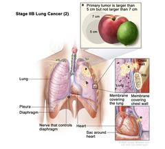 | Stage IIB lung cancer (2); drawing shows (a) a primary tumor (larger than 5 cm but not larger than 7 cm) in the left lung (top inset) and (b) a separate tumor in the same lobe of the lung as the primary tumor. Also shown is cancer that has spread to (c) the chest wall and the membranes covering the lung and chest wall (middle inset); (d) the nerve that controls the diaphragm; and (e) the sac around the heart (bottom inset). The pleura, diaphragm, heart, and a rib (middle inset) are also shown. |
06/26/2019 | |
 | Stage IIIC lung cancer; drawing shows a primary tumor in the left lung and (a) separate tumors in the same lobe of the lung with the primary tumor. Also shown is cancer in lymph nodes above the collarbone on the same side and opposite side of the chest as the primary tumor. Also shown is (b) cancer that has spread to the following: the chest wall and the lining of the chest wall and lung, the nerve that controls the voice box, the trachea, the carina, the esophagus, the breastbone, the diaphragm, the nerve that controls the diaphragm, the heart, the aorta and vena cava, and the sac around the heart. |
06/26/2019 | |
 | Stage IVA lung cancer; drawing shows a primary tumor in the right lung and (a) a tumor in the left lung. Also shown is (b) fluid or cancer nodules around the lungs or heart (inset), and (c) other organs or tissues where lung cancer may spread, including the brain, adrenal gland, kidney, liver, bone, and distant lymph nodes. |


|
06/25/2019 | |
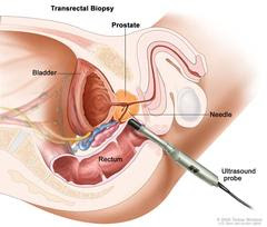 | Transrectal biopsy; drawing shows a side view of the prostate, bladder, and rectum. Drawing also shows an ultrasound probe with a needle inserted into the rectum to remove a tissue sample from the prostate. |
06/25/2019 | |
 | Anatomy of the digestive system; drawing shows the mouth, salivary glands, pharynx (throat), esophagus, stomach, liver, gallbladder, pancreas, small intestine, large intestine, rectum, and anus. |
06/25/2019 | |
 | Anatomy of the digestive tract; drawing shows the mouth, pharynx (throat), esophagus, stomach, small intestine, large intestine, rectum, and anus. |
06/25/2019 | |
 | Anatomy of the spine; drawing shows a side view of the spine, including the cervical spine (C1-C7), thoracic spine (T1-T12), lumbar spine (L1-L5), sacral spine (S1-S5), and the coccyx (tailbone). Also shown are the spinal cord, vertebra (back bone), conus medullaris (the end of the spinal cord), cauda equina (the bundle of spinal nerves that extend beyond the conus medullaris), and a lumbar disc. The clivus (a bone at the base of the skull near the spinal cord) is also shown. |
06/25/2019 | |
 | Drawing of a skeleton of a young girl. |
06/25/2019 | |
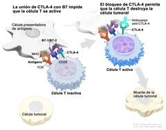 | Inhibidor de puntos de control inmunitario. En el panel izquierdo, se observa la unión del receptor de la célula T (TCR) con el antígeno y las proteínas del complejo principal de histocompatibilidad (MHC) en la célula presentadora de antígeno, y la unión de CD28 en la célula T con B7-1/B7-2 en la célula presentadora de antígeno. También se muestra la unión de B7-1/B7-2 con CTLA-4 en la célula T, que mantiene inactivas a las células T. En el panel derecho, se observa un inhibidor de puntos de control (anticuerpo anti-CTLA) que impide la unión de B7-1/B7-2 con CTLA-4, lo que permite que las células T se activen y destruyan las células tumorales. |
06/25/2019 | |
 | En la imagen de la izquierda se observa el sistema circulatorio en una mujer. En la imagen de la derecha se observa el sistema linfático en un varón. |
06/25/2019 | |
 | Cáncer de próstata en estadio I. Se observa una imagen con dos paneles. En el panel superior, se observa cáncer en menos de la mitad del lado derecho de la próstata que se detecta por una biopsia con aguja. En el panel inferior, se observa cáncer en menos de la mitad del lado izquierdo de la próstata que se detecta por un examen digital del recto. En ambos paneles, la concentración del PSA es menor de 10 y el grupo de grado es 1. También se muestran la vejiga, el recto y la uretra. |
06/25/2019 | |
 | Cáncer de próstata en estadio IIA. Se observa una imagen con dos paneles. En el panel superior, se observa cáncer en la mitad o menos de un lado de la próstata. La concentración del PSA es de 10 a menos de 20 y el grupo de grado es 1. En el panel inferior, se observa cáncer en más de la mitad de un lado de la próstata. La concentración del PSA es menor de 20 y el grupo de grado es 1. En ambos paneles, también se muestran la vejiga, el recto y la uretra. |
06/25/2019 | |
 | Cáncer de próstata en estadio IIB. En la imagen se observa cáncer en un lado de la próstata. La concentración del PSA es menor de 20 y el grupo de grado es 2. También se muestran la vejiga, el recto y la uretra. |
06/25/2019 | |
 | Cáncer de próstata en estadio IIC. En la imagen se observa cáncer en ambos lados de la próstata. La concentración del PSA es menor de 20 y el grupo de grado es 3 o 4. También se muestran la vejiga, el recto y la uretra. |
06/25/2019 | |
 | Cáncer de próstata en estadio IIIA. En la imagen se observa cáncer en un lado de la próstata. La concentración del PSA es de por lo menos 20 y el grupo de grado es 1, 2, 3 o 4. También se muestran la vejiga, el recto y la uretra. |
06/25/2019 | |
 | Cáncer de próstata en estadio IIIB. En la imagen se observa cáncer que se diseminó de la próstata a las vesículas seminales y al tejido cercano. El PSA es de cualquier concentración y el grupo de grado es 1, 2, 3 o 4. También se muestran la pared pélvica, la vejiga y el recto. |
06/25/2019 | |
 | Cáncer de próstata en estadio IIIC. En la imagen se observa cáncer en un lado de la próstata. El PSA es de cualquier concentración y el grupo de grado es 5. También se muestran la vejiga, el recto y la uretra. |
06/25/2019 | |
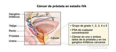 | Cáncer de próstata en estadio IVA. En la imagen se observa cáncer en un lado de la próstata y en los ganglios linfáticos cercanos. El PSA es de cualquier concentración y el grupo de grado es 1 ,2, 3, 4 o 5. También se muestran la vejiga, el recto y la uretra. |
06/25/2019 | |
 | Cáncer de próstata en estadio IVB. En la imagen se observan otras partes del cuerpo donde es posible que el cáncer de próstata se haya diseminado, como los ganglios linfáticos lejanos y el hueso. En un recuadro, se muestran células cancerosas que se diseminan desde la próstata, a través de la sangre y el sistema linfático, hasta otra parte del cuerpo donde se formó el cáncer metastásico. |
